
Vshaped wear in the cervical part of the tooth is known as dental
An understanding of this anatomy is essential for assessment and treatment of cervical spine problems. The cervical spine's major functions include supporting and cushioning loads to the head/neck while allowing for rotation, and protecting the spinal cord extending from the brain. [1] The cervical spine is subjected to extrinsic factors such.

Matriz Cervical TDV Restaurações classe V YouTube
The process of development of teeth is a very complex process resulting from interactions between the ectoderm of the oral cavity, which gives rise to cells that produce enamel, and the neural crest ectomesenchyme which gives rise to the tooth structures other than enamel. At first, i.e., during the six weeks of intrauterine life, the tooth germ starts growing, and the cells forming the.

SciELO Brasil Dental findings on face and neck imaging Dental
Cervical two is called the axis, as it is the one essential for allowing rotation to occur in the cervical spine. Both the atlas and axis are small, flat vertebrae. The atlas at cervical level one is a flat, ring-shaped vertebra. It does not have a vertebral body. The flat area on its superior surface supports your skull.

469 curtidas, 3 comentários 🦷Studygram de Odontologia 🦷
Invasive cervical resorption (ICR) is a relatively rare type of ERR (External Root Resorption), in which a localized resorption begins in the cervical area of the tooth, below the epithelial junction and above the ridge crest.. (RRE), no qual uma reabsorção localizada começa na região cervical do dente, abaixo da junção epitelial e.

Endodontia para dentes decíduos Blog Dental Speed
Cervical radiculopathy (also known as " pinched nerve ") is a condition that results in neurological dysfunction caused by compression and inflammation of any of the nerve roots of your cervical spine (neck). Neurological dysfunction can include radiating pain, muscle weakness and/or numbness. "Cervical" comes from the Latin word.
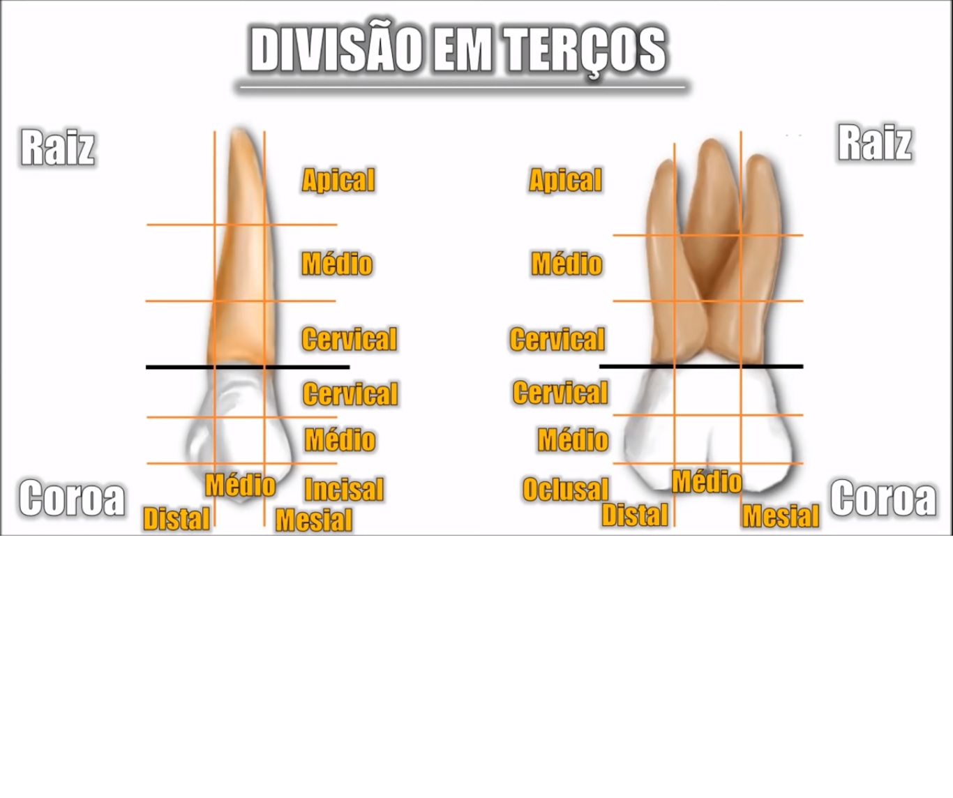
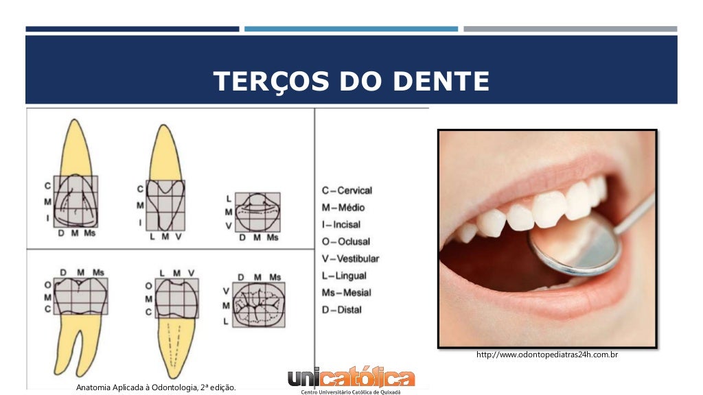
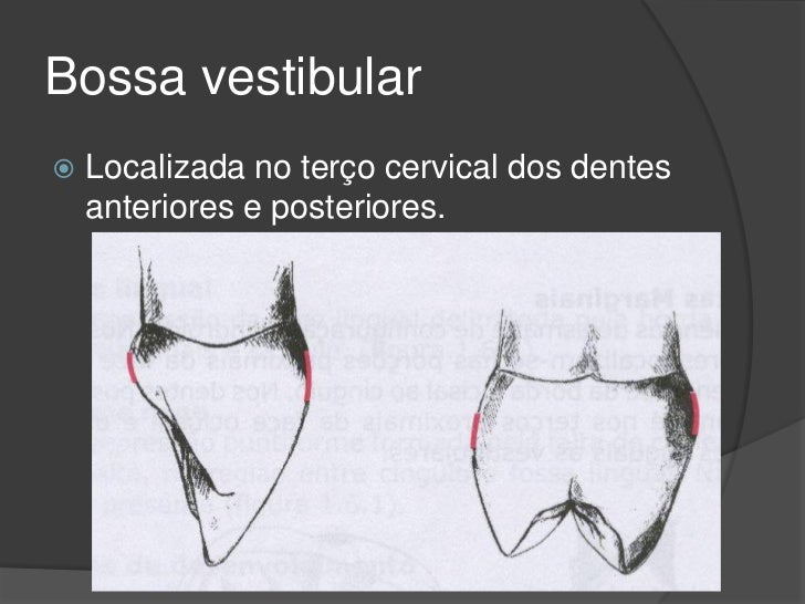
terços dos dentes Anatomia de Cabeça e Pescoço
Cervical Spine. The neck is part of a long flexible column, known as the spinal column or backbone, which extends through most of the body. The cervical spine (neck region) consists of seven bones ( C1-C7 vertebrae ), which are separated from one another by intervertebral discs. These discs allow the spine to move freely and act as shock.

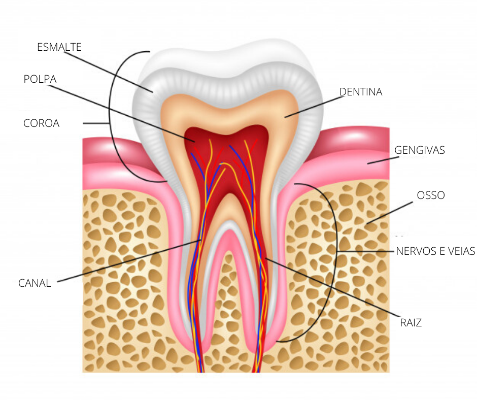
Anatomicamente a estrutura interna do dente pode ser dividida em
The spine, or vertebral column, is a segmental set of 33 bones and associated soft tissues in the subcranial portion of the axial skeleton. It is subdivided into 5 regions based on curvature and morphology: cervical, thoracic, lumbar, sacral, and coccygeal (see Image. Vertebral Column).

Aula de Anatomia Dental Incisivo Central Inferior Dente 31 e 41
O segmento cervical da coluna vertebral é muito importante dos pontos de vista anatômico e clínico. É nessa região que se originam os nervos dos braços, através do plexo braquial, e também é nela que o plexo cervical se origina para inervar o diafragma e outras estruturas.

anatomy location terms of teeth Introduction to Dental Anatomy
Introdução:: A reabsorção cervical invasiva (RCI) é um tipo relativamente raro de reabsorção radicular externa (RRE), no qual uma reabsorção localizada começa na região cervical do dente, abaixo da junção epitelial e acima da crista marginal. Objetivo:: Descrever o caso clínico de um menino com 11 anos de idade, sem histórico de trauma dentário, apresentando apinhamento.

Minuto anatômico 103 Faces e numerações dos dentes Anatomia
The cervical portion of the spine is an important one anatomically and clinically. It is within this region that the nerves to the arms arise via the brachial plexus, and where the cervical plexus forms providing innervation to the diaphragm among other structures. The cervical spine also allows passage of important vasculature to reach the brain and provides attachment sites for muscles that.
ANATOMIA DENTAL GENERALIDADES
Your seven cervical vertebrae (C1 to C7) are connected at the back of the bone by a type of joint (called facet joints), which allow for the forward, backward and twisting motions of your neck. Your cervical spine is also surrounded by muscles, nerves, tendons and ligaments.
Qual é a anatomia de um dente? Uniodonto Minas
The human vertebral column or spine has five distinct anatomical regions: cervical, thoracic, lumbar, sacral, and coccygeal. However, the cervical spine is a potential area of importance due to its proximity to the head, containment of the upper spinal cord, and vertebral arteries that contribute to the posterior circulation of the brain. Seven cervical vertebrae, combined with cartilages.
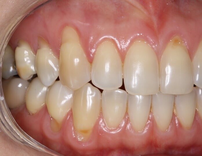
Cárie ou lesões cervicais não cariosas?
Anatomia do dente O dente é uma das estruturas anatômicas e histológicas mais individuais e complexas do corpo. A composição tecidual do dente só é encontrada no interior da cavidade oral, e é limitada às estruturas dentárias.
Revisão anatômica dental
Na vertical: terço oclusal/incisal, terço médio, terço cervical. Essa subdivisão é feita para que a identificação seja precisa. Por exemplo, se o dentista identificar uma cárie na face vestibular, pode ser em qualquer parte dessa face.

Linha Equatorial Do Dente ENSINO
Summary. Degenerative cervical spine disease (cervical spondylosis) is osteoarthritis of the spine, which includes the spontaneous degeneration of either disk or facet joints. Presenting symptoms include axial neck pain and neurologic complications. The most common neurologic complication is cervical spondylotic radiculopathy.
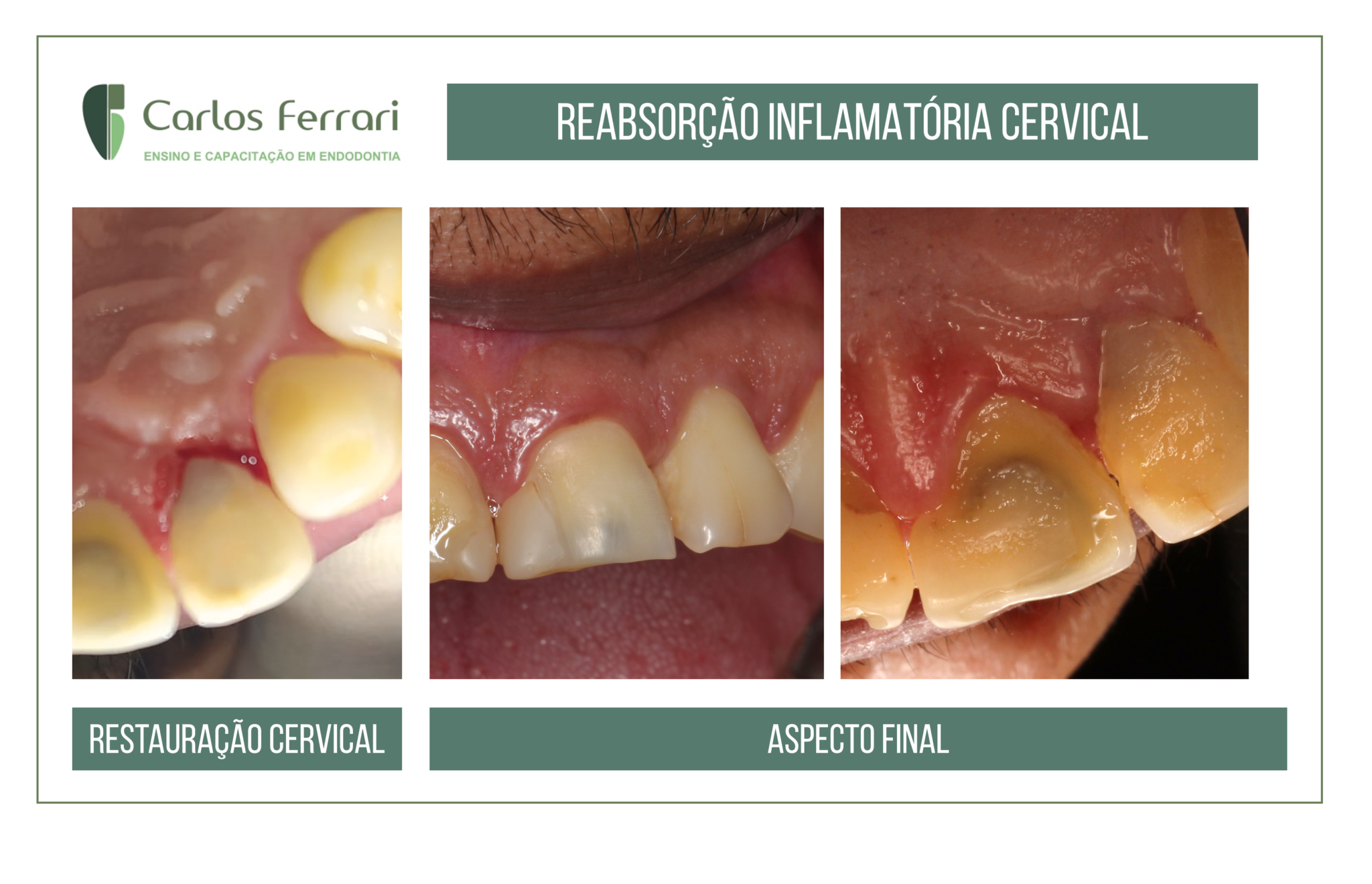
Reabsorção cervical externa. Tratamento endodôntico e cirúrgico
Classification. There are two classification systems 5,6. Anderson and D'Alonzo. most commonly used, describes level of fracture line (i.e. through tip, base, or lateral masses) Type I: Avulsion fracture of the upper dens; usually stable. Type II: Transverse fracture of the base of dens; unstable. Type III: Fracture at the body of C2.
